Home
/ Back Muscles Anatomy : Back Muscles Doccheck - Three types of back muscles that help the spine function are extensors, flexors and obliques.
Back Muscles Anatomy : Back Muscles Doccheck - Three types of back muscles that help the spine function are extensors, flexors and obliques.
Back Muscles Anatomy : Back Muscles Doccheck - Three types of back muscles that help the spine function are extensors, flexors and obliques.. Back pain is one of the most common kinds of pain for adults, and muscle strains are the most common type of back pain. This blog post article is an overview of the muscles of the lumbar spine of the trunk. On this page, you'll learn about each of these muscles, their locations and functional anatomy. Back muscles, functions and exercises: Human musculature bodybuilding infographic muscular system vector human anatomy back muscle anatomy bicep male muscular anatomy human body anatomy female female anatomy muscle hamstrings muscle.
These are the muscles that are farther from the surface, closer to the internal organs and the spine. These layers of back muscles help to mobilize and stabilize your trunk during your day to day activities. This curve, called lordosis, helps to: These muscles give height and breadth to back development. The muscles, bones, ligaments, and tendons in the back can all be injured and cause back pain.
Muscles Move And Support The Spine from cloud2.spineuniverse.com The back consists of the spine, spinal cord, muscles, ligaments, and nerves. These muscles include the large paired muscles in the lower back, called erector spinae, which help hold up the spine, and gluteal muscles. Able to move the upper limb as they originate at the vertebral column and insert onto either the clavicle, scapula or humerus. The extensor muscles are attached to back of the spine and enable standing and lifting objects. (2017, elsevier) should be consulted. We think this is the most useful anatomy picture that you need. Anatomynote.com found anatomy of back muscles diagram from plenty of anatomical pictures on the internet. Human musculature bodybuilding infographic muscular system vector human anatomy back muscle anatomy bicep male muscular anatomy human body anatomy female female anatomy muscle hamstrings muscle.
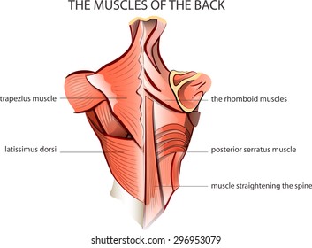
The surface muscles of the upper back include the trapezius muscles (traps) and posterior deltoids.
Includes latissimus dorsi, the trapezius, levator scapulae and the rhomboids. The intrinsic back muscles are found deeper to the extrinsic muscles, separated from them by the thoracolumbar fascia. Balance the weight of your head on top of your spine evenly distribute weights from your upper body into the lower extremities Muscle origin insertion action innervation artery notes; See back muscle anatomy stock video clips. This curve, called lordosis, helps to: Muscle and bone anatomy 12 photos of the muscle and bone anatomy back muscles and bones anatomy, human muscle and bone anatomy, muscle & bone anatomy 3d free download, muscle and bone anatomy app, muscle and bone anatomy quiz, human muscles, back muscles and bones anatomy, human muscle and bone anatomy. Both the deltoid and the trapezius are firmly attached to the spine of the scapula. What are the lower back muscles and their anatomy? Leaning back to straight vertical and all points in between. Anatomy of back muscles your back consists of three distinct layers of muscles, namely the superficial layer, the intermediate layer, and the deep layer. Three types of back muscles that help the spine function are extensors, flexors and obliques. Able to move the upper limb as they originate at the vertebral column and insert onto either the clavicle, scapula or humerus.
The extrinsic back muscles are located in the back, but act to produce movements of the shoulder and assist respiration. To perform clinical clinical orthopedic manual therapy to the lumbar spine. Back pain is one of the most common kinds of pain for adults, and muscle strains are the most common type of back pain. Related posts of muscles of the lower back and hip diagram muscle and bone anatomy. 1 your spine in this region has a natural inward curve.
Back Muscle Anatomy Diagram Quizlet from o.quizlet.com The trapezius and latissimus dorsi muscles connect the upper limb to the vertebral column. These structures work together to support the body, enable a range of movements, and send messages from the brain to the. The muscles of the back can be arranged into 3 categories based on their location: Extends and laterally bends the neck and head, rotates head to the same side: Human musculature bodybuilding infographic muscular system vector human anatomy back muscle anatomy bicep male muscular anatomy human body anatomy female female anatomy muscle hamstrings muscle. They start at the top of the neck and go down to the tailbone. The muscles of the back muscles make up a large part of the anatomy (structure) of the back. (2017, elsevier) should be consulted.
Muscle and bone anatomy 12 photos of the muscle and bone anatomy back muscles and bones anatomy, human muscle and bone anatomy, muscle & bone anatomy 3d free download, muscle and bone anatomy app, muscle and bone anatomy quiz, human muscles, back muscles and bones anatomy, human muscle and bone anatomy.
To control the posture of the entire body. The muscles of the back can be arranged into 3 categories based on their location: For more anatomy content please follow us and visit our website: We hope this picture anatomy of back muscles diagram can help you study and research. Includes latissimus dorsi, the trapezius, levator scapulae and the rhomboids. Balance the weight of your head on top of your spine evenly distribute weights from your upper body into the lower extremities The superficial back muscles are situated underneath the skin and superficial fascia. The back muscles are anatomically layered into superficial (extrinsic) and deep (intrinsic) muscles. The extrinsic back muscles are located in the back, but act to produce movements of the shoulder and assist respiration. To perform clinical clinical orthopedic manual therapy to the lumbar spine. These muscles give height and breadth to back development. The muscles, bones, ligaments, and tendons in the back can all be injured and cause back pain. Your lower back (lumbar spine) is the anatomic region between your lowest rib and the upper part of the buttock.
The muscles of the lower back, including the erector spinae and quadratus lumborum muscles, contract to extend and laterally bend the vertebral column. Muscle origin insertion action innervation artery notes; (2017, elsevier) should be consulted. Your lower back (lumbar spine) is the anatomic region between your lowest rib and the upper part of the buttock. Leaning back to straight vertical and all points in between.
Back Muscle Anatomy Images Stock Photos Vectors Shutterstock from image.shutterstock.com The back consists of the spine, spinal cord, muscles, ligaments, and nerves. The back muscles are anatomically layered into superficial (extrinsic) and deep (intrinsic) muscles. The superficial back muscles are situated underneath the skin and superficial fascia. The lower back (where most back pain occurs) includes the five vertebrae in the lumbar region and supports much of the weight of the upper body. Muscle origin insertion action innervation artery notes; The muscles, bones, ligaments, and tendons in the back can all be injured and cause back pain. These layers of back muscles help to mobilize and stabilize your trunk during your day to day activities. Back pain is common and might be caused by a problem with a muscle.
They start at the top of the neck and go down to the tailbone.
For more anatomy content please follow us and visit our website: As a general group, they extend from the neck to the sacrum and fulfill a basic and fundamental function: These muscles provide posture and stability to the body by holding the vertebral column erect and adjusting the position of the body to maintain balance. These are the muscles that are farther from the surface, closer to the internal organs and the spine. Understanding lower back anatomy is key to understanding the root of lower back and hip pain. The spaces between the vertebrae are maintained by intervertebral discs that act like shock absorbers throughout the spinal column to cushion the bones as the body moves. The back consists of the spine, spinal cord, muscles, ligaments, and nerves. The back muscles are anatomically layered into superficial (extrinsic) and deep (intrinsic) muscles. These muscles give height and breadth to back development. The intrinsic back muscles are found deeper to the extrinsic muscles, separated from them by the thoracolumbar fascia. The lower back (where most back pain occurs) includes the five vertebrae in the lumbar region and supports much of the weight of the upper body. The muscles of the back can be arranged into 3 categories based on their location: Able to move the upper limb as they originate at the vertebral column and insert onto either the clavicle, scapula or humerus.